Our Research

Mechanisms underlying cell identity and plasticity

Our lab is interested in delineating the lineage relationships of fibroblast subsets and their contribution to lung function or repair after injury. Depending on the organ and tissue type, under homeostatic or steady-state conditions, some cells can proliferate and differentiate into other specialized cell types. In barrier organs, such as the skin, gut and lung, cells exposed to the external environment undergo regular cycles of proliferation and differentiation to maintain a functional barrier. In the lung, the epithelial layer is in direct contact with inhaled air. Research efforts over the last two decades have carefully mapped out the replicative nature of sub-types of epithelial cells and pathways that are activated to promote differentiation.

Recent studies have shifted attention to other cell types in the lung, namely endothelial cells and fibroblasts/smooth muscle. Fibroblasts and smooth muscle surround the epithelium and vasculature in the lung. 
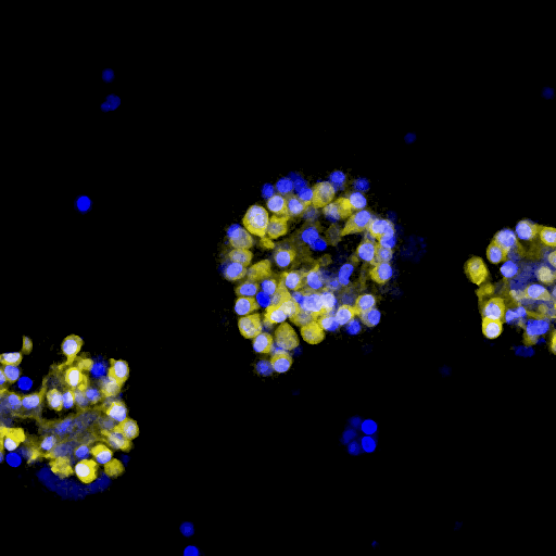
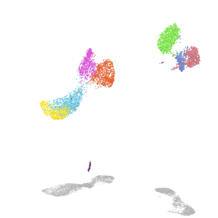
Using a combination of mouse genetic lineage tracing, fluorescence imaging and single-cell RNA sequencing, functionally distinct subsets of fibroblasts are being characterized. These fibroblasts have unique capacities to proliferate, differentiate and secrete growth factors that support epithelial or endothelial cell growth. We are intent on identifying signaling pathways and transcription factors that control the differentiation of fibroblasts.

lung mesenchyme scRNA, courtesy R. Windmueller

Mechanisms of alveolar development

Our lab is testing how mesenchymal lineages contribute to lung development using mouse genetics and single-cell RNA/ATAC-sequencing. Moreover, we are actively developing new assays to parse the varied functions of these mesenchymal cell subsets including migration and force-exertion assays. Lastly, we are exploring how co-development of mesenchymal and immune lineages occur at the onset of environmental exposure.

The alveolus is where the defining feature of the lung, gas-exchange, occurs. The alveolar compartment develops in a period that spans the transition to air breathing at birth. During this developmental stage, known as alveologenesis, the alveolar compartment undergoes morphogenic processes that expand the overall epithelial surface area by increasing the total number of alveoli. While there are likely multiple cellular sources that contribute to this morphogenic process, the more striking observation is a fibroblast lineage, that expresses smooth muscle actin (Acta2), referred to as a secondary crest myofibroblast (SCMF). This cell type is transient, existing exclusively during this time period, afterwards most of these cells are lost through apoptosis although some appear to remain. These data underscore the dynamic states and functions of the alveolar mesenchyme.

Alveolar Septa Formation

Translational impact for lung physiology and disease

Guiding our research is that the pulmonary system, as a whole, performs the essential function of gas-exchange. Our approach is to uncover mechanisms regulating unique cellular processes and then determine the impact of these processes on lung function or disease onset/progression. We use physiologic parameters to assess outcomes in our murine genetic models. We employ in vivo models that provoke cell behaviors in settings of tissue stress. Moreover, we have collaborations across the CHOP and PENN community to obtain clinical samples and primary cells to validate novel markers and cell phenotypes.

For example we can model cell interactions using organotypic three-dimensional tissue culture by mixing different cell populations and assessing cell growth or transcriptomic signatures. Further, we are expanding upon our novel murine reporters to generate human cell lines using gene-editing. These lines are being evaluated using our cell-based phenotyping assays for target discovery.

Alveolar organoid